

It can be stated that neoplasms found in the cephalic and cervical regions are rare, given their incidence of only 3%, when compared to other malignant cancers that affect humans. Within this statistic, squamous cell carcinoma (SCC) stands out as the most common histological class. Such tumors, although uncommon, are of great clinical importance and their severity is closely related to the tumor stage at the time of diagnosis. Furthermore, it is worth highlighting that the location of the mass is of great importance, as it is a region made up of numerous noble structures, making surgical procedures unfeasible as a treatment option, given that there is extensive anatomical complexity. This research is characterized by being a case report of anatomical and histopathological findings of SCC (squamous cell carcinoma) in the right nasal cavity of a cadaver, with undetermined name, located at the Anatomical Center I of UNIFENAS, Alfenas campus.
Keywords: Nasal cavity tumor, Invasive squamous cell carcinoma, Squamous cell carcinoma
Every anatomy student, when faced with a cadaver for their practical studies, also comes across feelings of respect and empathy for the person who is providing their body for study. However, much information such as name, age, life history, cause of death and how that cadaver ended up on that table is generally unknown to the students. Such information is only obtained not only through the anatomical study itself but through dissection techniques that explore every detail, making it possible to detect abnormalities in the inert body that were not seen before and that help to elucidate the cause of death.
Among several findings that we can find, in this case an epidermoid carcinoma was found in the cephalic region with a focus on the right nasal cavity. Epidermoid carcinoma, also called squamous cell carcinoma, is a malignant tumor that affects the keratinized cells of the epidermis and invades the dermis, which can lead to large affected areas or, in more advanced stages, metastasis. Its main etiology is related to prolonged exposure to ultraviolet radiation, which causes mutations and immunosuppressive effects essential in the processes of light-induced carcinogenesis. The p53 protein suppressor gene stands out, as it plays an important role in supervising the genetic material and eliminating cells that have undergone mutations through the mechanism of apoptosis of the abnormal cell.1,2
Neoplasms in the head and neck region are relatively rare, accounting for approximately 3% of all cancers. The most common malignant tumors that affect the nasal cavity are squamous cell carcinoma, melanoma, adenoid cystic carcinoma, sarcoma, adenocarcinoma, neuroendocrine carcinoma, undifferentiated carcinoma, non-Hodgkin's lymphoma and esthesioneuroblastoma. Epithelial tumors, such as squamous cell carcinoma, predominate. However, due to their anatomical location, these tumors are difficult to treat surgically.3
Squamous cell carcinoma is the second most common type of skin cancer after basal cell cancer, with more than 1 million cases in the United States and 2,500 deaths per year. In Europe, the incidence ranges from 9 to 96 per 100,000 inhabitants in the male population and from 5 to 68 per 100,000 inhabitants in the female population. In Brazil, in 2014, approximately 576,580 new cases of squamous cell cancer were reported in the oral region alone.1,4
In 2019, according to data from INCA (National Cancer Institute – Brazil), there were 20,722 deaths from head and neck cancer. The head of the INCA Head and Neck Section, surgeon Fernando Dias, explains that the high mortality rate is mainly due to patients arriving for treatment with the disease already advanced. When the diagnosis is made in the early stages, the patient has, on average, an 80% chance of survival.5
In most cases, nasal cavity and paranasal sinus cancers are found because of the problems they cause. Finding these cancers in people without symptoms is rare and usually by accident (during tests to check for other medical problems). Possible signs and symptoms of these cancers (usually on one side only) include: Nasal congestion that does not improve or gets worse; Pain above or below the eyes; Blockage on one side of the nose; Postnasal discharge (nasal drainage in the back of the nose and throat); Nosebleeds; Pus draining from the nose; Problems with smell; Numbness or pain in parts of the face; Looseness or numbness of the teeth; Lump or mass on the face, palate (top of the mouth), or inside the nose; Constant watery eyes; Bulging of one eye; Loss or change in vision; Pain or pressure in one ear; Hearing loss; Headache; Difficulty opening the mouth; Swollen lymph nodes in the neck. Having one or more of these symptoms does not mean you have nasal cavity or sinus cancer.6
The purpose of this report is to identify the anatomical-histopathological samples of the tissues found from the corpse described below, as well as to report its incidence in the general population, and the referenced treatment.
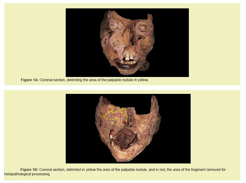
Donated human body preserved in 3% formaldehyde solution, approximately 50 years old, male, Afro-descendant, undetermined name, was subjected to a coronal cut on the head for the purpose of studying its cavities, carried out on an industrial band saw owned by the company. anatomical center. However, a mass in the nasal cavity was evident, being a palpable nodule, an investigation was suggested. An incisional biopsy of approximately 1 cm was performed on this mass, and it was submitted to the pathology service of UNIFENAS (Alfenas) for histopathological treatment in H&E and respective analysis for issuing a technical report (Figures 1A and 1B).
The risks were considered minimal, as the head cut was performed by the professor responsible for the laboratory (Prof Dr Luis Henrique Rapucci Moraes), taking due biosafety precautions.
Macroscopic results
According to the technical report from the Pathology laboratory at Unifenas, the material was received in 10% formalin and consisted of a portion of irregular, yellowish-brown tissue measuring 1.0 x 0.8 x 0.7 cm, which was elastic and homogeneous when cut.
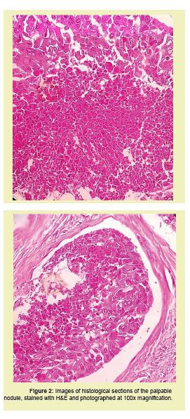
All material was subjected to histological examination with H&E (2fs/1bl).
The images were captured using a camera attached to a Nikon binocular microscope (E100) at 100x magnification Figure 2.
Microscopic Results
The result of the histopathological examination was analyzed by Prof. Dr. Fiorita Gonzales Lopes Mundim, who described the following finding
Moderately differentiated invasive squamous cell carcinoma with areas of autolysis and the following characteristics
• Type of resection: incisional biopsy• Histological grade: g2/moderately differentiated
• Level of invasion: the neoplasm infiltrates the nasal mucous glands up to the muscular plane in the cuts made
• Perineural invasion: not detected
• Angiolymphatic invasion: not detected
• Ulceration: not detected
• Surgical margins: deep margin compromised by the neoplasm
Malignant tumors of the nasal cavity and paranasal sinuses represent only 3% of all head and neck carcinomas. Within this subgroup, squamous cell carcinoma (SCC) is the most common cancer of the nasal cavity.3
SCC of the nasal cavity is a rare tumor entity due to the lack of information, as it is generally studied together with paranasal and sinus carcinomas, as well as other malignant entities. The overall incidence of sinonasal cancer has been considered, worldwide, with an incidence of 0.556 cases per 100,000 inhabitants, compared to more common head and neck cancers (e.g., pharynx, larynx or oral cavity) with an incidence of 1 to 5 per 100,000 inhabitants. The most common malignant sinonasal tumor is SCC, which represents 50% of all cases.7
The management of these tumors is often challenging because of their local aggressiveness, tendency to grow rapidly, and particularly the devastating consequences and psychosocial sequelae of aggressive facial surgery. Nasal cavity tumors are usually rare, so they are analyzed together with other tumors of the nasal cavity, paranasal sinuses, or nasal vestibule. This generates non-homogeneous results because of the different histological types and stages. In addition, nasal cavity tumors have a substantially better survival rate compared to paranasal sinus cancers.3
As we can see in the images in Figure 1, there was cellular invasion into the right maxillary sinus, which contributes to greater severity of the disease and complexity of treatment.
Potential risk factors for SCC of the nasal cavity reported in the literature include smoking and alcoholism, and are compromised by the interaction of environmental factors and genetic inheritance for the development of the disease.8 Also, in recent years, the human papillomavirus (HPV) has been shown to be an important causative agent in some head and neck carcinomas, especially in carcinomas of the oral cavity and oropharynx. The role of HPV in SCC of the nasal cavity is still unclear; they appear to be biologically relevant in laryngeal carcinogenesis.9
We were unable to know in advance whether the corpse investigated was a smoker, but we could notice his lungs with numerous dark spots, a characteristic color caused by carbon monoxide.
Treatment for SCC
After someone is diagnosed with nasal cavity or sinus cancer, doctors will try to find out if it has spread and, if so, how far. This process is called staging. The stage of a cancer describes how much cancer is present in the body. It helps determine how serious the cancer is and how best to treat it.6
Chemotherapy for nasal cavity and sinus cancer may involve a combination of several drugs. These cancers are rare, so there aren’t many studies to help doctors decide how best to treat them. Doctors often treat them with the same drugs they use for other, more common head and neck cancers. The most common are squamous cell carcinoma, adenocarcinoma, and adenoid cystic carcinoma—all of which can be grouped together as carcinomas.6,10-16
Some of the chemotherapy drugs most commonly used to treat carcinomas, according to the American Cancer Society, include:
• Cisplatin
• Carboplatin
• 5-fluorouracil (5-FU)
• Docetaxel (Taxotere)
• Paclitaxel (Taxol)
• Methotrexate
• Capecitabine (Xeloda), a pill that is converted to 5-FU when it reaches the tumor
The drugs used depend on many factors, including the type of cancer, the extent of the cancer, the person's overall health, and whether chemotherapy is combined with radiation therapy. The chemotherapy drugs used first include cisplatin or carboplatin alone or in combination with 5-FU, but other combinations are also available. For people who cannot tolerate chemotherapy, the targeted therapy drug cetuximab is often used instead of radiation.6,17-23
Therefore, it can be said that Squamous Cell Carcinoma of the Nasal Cavity, despite its rare incidence, has great clinical importance, given its location, in relation to its proximity to noble structures, in addition to its metastatic and invasive characteristics, as noted in this study. Such factors, in this case, corroborate a possible cause of death of the individual. Here is our contribution in exposing this pathology directly positioned on the cadaver, where such images can contribute to educational-scientific enrichment in the area of human health.
None.
This Case Report received no external funding.
Regarding the publication of this article, the authors declare that they have no conflict of interest.
