Cardiac tamponadeis a medical emergency which requires a fast diagnosis and treatment. We report the successful management of 51-year-old women who presented with cardiac tamponade due to ventricular rupture. Once this condition was suspected and confirmed by echocardiography, an emergent pericardiotomy was made. This case highlights the importance of a prompt diagnosis and how this could change the prognosis.
Glossary: AMI: Acute Myocardial Infarction, EKG: Electrocardiogram, ICU, Intensive Care Unit, NIRS: Near-infrared Spectroscopy, SctO2: Cerebral Oxygen Saturation, TEE:Transoesophageal Echocardiogram, TTE: Transthoracic Echocardiography
Cardiac tamponade is caused by an abnormal increase in fluid accumulation in the pericardium, which impedes normal cardiac filling and reduces cardiac output.1 This entity can develop in patients with any condition that affects the pericardium. A high index of suspicion can decrease concomitant morbidity and mortality. We present a case of an acute cardiac tamponade secondary to ventricular rupture after a myocardial infarction. This complication is associated to a high mortality rate and prompt diagnosis and treatment can be lifesaving.
A 51-year-old Caucasian woman, with hash moto thyroiditis and dyslipidaemia, presented to the emergency department of a tertiary hospital. She had an appointment at her primary care doctor 15days earlier due to an epigastric pain and vomiting. Upon arrival, the patient was in a state of hemodynamic collapse. She was lethargic with an invasive blood pressure of 65/35mmHg and a pulse rate of 110beats/min. On physical examination there were muffled heart sounds.
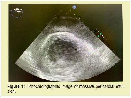
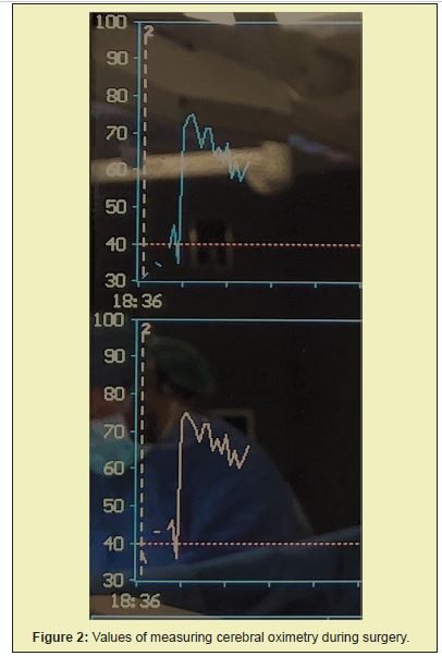
ST segment elevation and Q Waves in the anterior leads are evident on the electrocardiogram (EKG). Transthoracic echocardiography (TTE) revealed a severe depression of the left ventricular systolic function, an apical aneurysm with a distal rupture of the posterior ventricular wall and a massive pericardial effusion compromising right ventricular filling (Figure 1). Vasopressor support was initiated. The case was then discussed with the cardiothoracic surgeons and the anaesthetic team of the central hospital of reference who decided to admit the patient for an emergent pericardiotomy. Immediately upon arrival at the operating room, standard preoperative monitoring was applied along with near-infrared spectroscopy (NIRS). The induction was performed with iv propofol 30 mg, fentanyl 50 mcg and rocuronium 50 mg. Once the incision was made, blood and a giant clot were seen within the pericardial space (supplemental video 1). There was sudden hemodynamic recovery following clot removal with a pronounced improvement of arterial pressure. Furthermore, the baseline non-invasive cerebral oxygen saturation (SctO2) levels of 45% and 40% for the left and right sides of the brain respectively, immediately increased at this time to around 75% bilaterally (Figure 2), showing rapid recovery of cerebral perfusion pressure and therefore resolving the superior vena cava syndrome secondary to the cardiac tamponade.


Video 1: Intra-operative video of the giant clot within the pericardial space.
Due the consumptive coagulation prothrombin complex was administered to restore effective hemotasis.
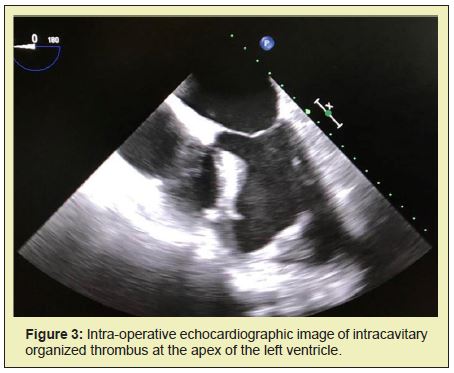
Intraoperative transoesophageal echocardiogram (TEE)also showed an intracavitary organized thrombus at the apex of the left ventricle that was left untouched (Figure 3), to be posteriorly treated conservatively with anticoagulation. The patient was transported to the intensive care unit (ICU) in a stable condition, was extubated on post-operative day two and discharged on day eleven.

Ventricular rupture is the more severe complication of acute myocardial infarction (AMI) and is more common in women and the elderly.2 A successful outcome demands early recognition of the presenting clinical signs coupled with rapid confirmation of the diagnosis, ideally by echocardiography, andprompt appropriate treatment. Coagulopathies and cardiac tamponades are two of the major reasons for an emergent run to the operating room. For optimal outcomes, management must be carefully coordinated between the anaesthesiologists and the cardiac surgical team. Many patients can be admitted to the operating room on an emergency basis, arriving in a severely compromised hemodynamic state.2 Thus, initial management is to restore some form of hemodynamic stability and the induction of anaesthesia should be accomplished with minimal compromise in cardiac function.2
In the presence of copious surgical drainage, supportive treatment should go hand in hand with surgery to obtain a better result. As a conclusion, this case illustrates the challenges that all medical teams are confronted with when dealing with a patient with left ventricular rupture, which requires an extensive pathophysiologic knowledge and careful hemodynamic control.3 Furthermore, it was also interesting to show a superior vena cava syndrome secondary to cardiac tamponade, which was immediately resolved and documented through non-invasive cerebral oxygen saturation levels.
None.
None.
Author declares that there is no conflict of interest.
- 1. Appleton C, Gillam L, Koulogiannis K. CardiacTamponade. Cardiology Clinics. 2017.
- 2. Bard J. Anesthetic implications of subacute left ventricular rupture following acute myocardial infarction: A case report. AANA Journal. 2000.
- 3. O’Connor C, Tuman K. The Intraoperative Management of Patients with Pericardial Tamponade. Anaesthesiology Clinics. 2010;28(1):87‒96.

