Open rhinoplasty techniques as well endonasal techniques that involve lower lateral cartilage delivery and marginal incisions lead to many unpleasant effects such as alar notching, alar retraction, alar collapse, dropped/pinched tip, Pollybeak, asymmetry, alar collapse, deviation, and dimpling. Both the original Joseph endonasal technique and the later Goldman endonasal tip technique has also resulted in the above issues, while these problems are well documented in the open rhinoplasty approach. To reduce these sequalae, the author has developed a new combination technique. It is based on the Goldman's tip and the I-beam of intermediate crura while avoiding lateral crus marginal incision and delivery (which are the culprits associated with a poor outcome). This results in an intact alar rim reducing healing problems along the alar rim. Furthermore, lateral crus preservation together with a strong tripod of conjoined lateral to medial crura supported by a columellar strut and tip grafts lead to significantly enhanced tip support thus substantially reducing negative sequalae such as asymmetry, alar collapse, dropped tip, pinching, notching and alar retraction while at the same time making the outcome more predictable. The author has been applying this technique since 1999 (Last 23 years). The technique and follow-up will be fully discussed.
Keywords: Rhinoplasty, Goldman's tip, Modified oblique dome division
Exposure of the lateral crus via the open approach or delivery of the lateral crus in the endonasal approach are associated with many of the unpleasant sequalae following rhinoplasty. The nose is a hollow organ with thin walls, therefore, any mild degree of wound contracture, scarring or fibrosis (which is most cases is inevitable) may lead to unpleasant effects such as alar retraction, alar notching, alar collapse, dropped tip, asymmetry, Pollybeak deformity, pinched tip and so on. The principal basis of our preservation technique, referred to as “Modified Oblique Dome Division” is to fully preserve the lateral crus. The author recommends this technique following an experience of over 20 years and over several thousand cases. This technique is particularly useful to achieve tip projection, symmetry, definition, and refinement in the following situations:1-5
- Tip Under-projection
- Tip Over-projection
- Short columella
- Supra-alar fullness
- Tip asymmetry
- In revision cases when lateral crus delivery is technically difficult
The principles are:
- Modified Oblique Dome Division
- Intermediate Crus Delivery and I-beam
- No Lateral Crus delivery
- No lateral crus marginal incision
- Creation of strong cartilaginous foundation in the midline of conjoined medial to intermediate crura supported by Columella strut and as needed additional Columellar-Tip graft.
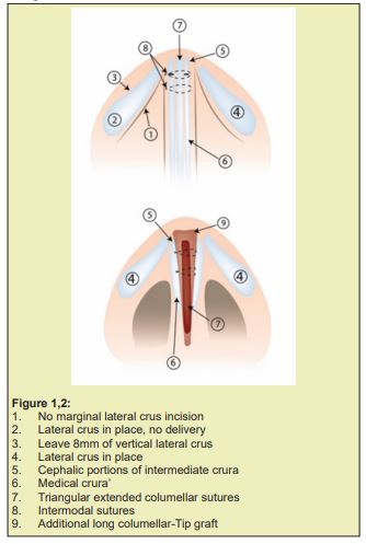
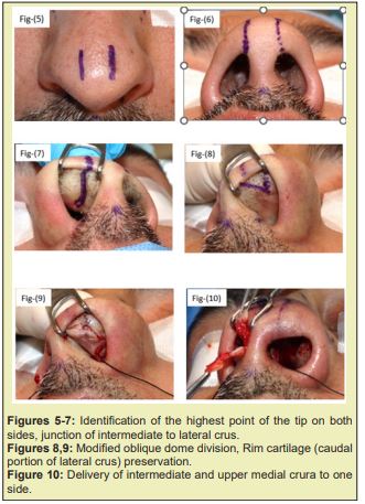
- Mark the tip defining point on both side of the tip (highest point on the tip). Next, mark it down on the alar rim, and then internally on the alar cartilage. The line joining these two points will separate lateral crus from the intermediate crus Figures 1-7.
- The procedure is usually done with local anaesthesia with sedation although if the surgeons prefers, it can be done under general anaesthetic.
- An Endonasal approach and obtaining septal or conchal graft.
- Marginal columella incision from mid-columella and up to the level of the external soft triangle at junction of intermediate to lateral crura. There is no marginal incision along the caudal margin of the lateral crura.
- Modified Oblique dome division:
- The incision is now extended obliquely in the intermediate crus from medial to lateral. In a way to fully preserve the rim cartilage and divide the intermediate crura into the caudal portion (Rim cartilage) and a Cephalic portion (Never borrow or steal any portion of lateral crus) Figures 8,9.
- The bilateral Cephalic portion of intermediate crura are delivered to one side. (Usually right side) Figures 10.
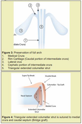
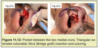
- A pocket is created between the medial crura down to nasal spine and caudally to expose the caudal septum, and a Triangular Extended Columellar Strut is now inserted and sutured (5/0 PDS) to both medial crura and Caudal septum. (Bridging between the medial crura and caudal septum). This will obtain a strong sustained tip projection, elevation, and rotation Figures 11,12.
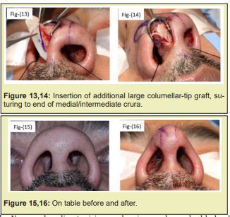
- An additional Tip-Columellar extended graft is positioned and sutured to the caudal margin of the intermediate and medial crus. The grafts should be crushed and bevelled in order to avoid demarcation. The graft is long in order to augment the Naso-labial angle and obtain more rotation Figures 13,14.
- The re-oriented complex structure of medial and intermediate crura with columellar strut and the tip graft are pushed back inside the nose into a normal position. The transfixion and marginal incisions are closed with 5/0 PDS. Then two additional septocolumellar sutures are used to fix the medial crura/ columellar strut complex to the caudal septum Figures 15,16.
- Now under direct vision and using a sharp double-hook, 1-2mm of cephalic lateral crus is trimmed to reduce supra-alar fullness and allow space for rotation.
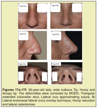
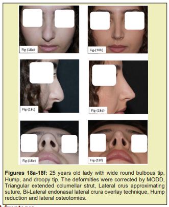
- The cartilaginous foundation in the midline of conjoined medial crura to the intermediate crura, extended triangular columellar strut, additional columella and tip graft together with septocolumellar sutures and footplate sutures establishes a very strong foundation that provides enhanced tip projection, symmetry, definition, refinement and rotation with full preservation of the integrity of the lateral crus (No marginal incision or delivery) Figures 17,18.
- Preservation of the entire lateral crus on the alar sidewalls thus reducing alar retraction, notching, collapse, dimpling, asymmetry and even deviation.
- The trimming of cephalic lateral crus is made more visible, more accurate and has better control.
- Less postoperative oedema and crusting.
- More natural looking alar sidewalls.
- Stretches the alar sidewalls and reduces the alar flare.
- Achieves enhanced tip projection, elevation, definition, rotation, and refinement.
- Corrects mild tip bulbosity, when combined with conservative retrograde cephalic trimming of the lateral crura (ten mm to be preserved) and combined with lateral crus approximating suture.
- Corrects over projecting tip. This is achieved by conservative trimming of the proximal ends of the lateral crura and superior ends of the intermediate crura.
- The author believes that lateral and medial crura overlay technique is best for correction of the over projected tip as you will have direct control.
- It should be performed in a very precise, accurate and symmetrical manner. Otherwise, unilateral, or bilateral alar collapse, pinching, notching or retraction might occur, which will require revision with Batten grafts. Anyone unwilling to work carefully to attain a stable symmetrical structure should not attempt this technique (Never borrow or steal any portion of lateral crus).
- Thin skin patients may develop mild tip pinching, therefore avoid lateral crus approximating suture in patient with thin.
The Lateral Crus Approximating Suture is extremely useful in almost 90% of the cases except in patients with very thin skin. Using 5/0 PDS or 6/0 Proline, the suture is passed through superior septal angle from left to right, then through the cephalic lateral crus on the right side, then back through the cartilaginous dorsum 1mm behind SSA and then passed through the left lateral crus cephalic margin and tied. This suture is of great utility for the following reasons:
- It results in supra-alar fullness with more tip definition and refinement, it stabilizes the bi-lateral crus to the cartilaginous dorsum portion.
- It stretches the alar sidewalls and reduces alar flare.
- It stabilizes the midline cartilaginous foundation (of the Medial crura, Intermediate lateral crus, Columellar strut and Tip graft) and increases tip rotation.6-10
The author from years 1991 to1999 was mainly performing open rhinoplasty and endonasal delivery approach in which the lateral crus were fully exposed or delivered respectively. The author was practicing in the Middle East & Gulf Regions on mainly non-Caucasian patients. Using the Open approach & Delivery approach in this part of the world resulted in many unpleasant problems, such as notching, alar retraction, alar collapse, tip asymmetry, nostrils asymmetry and even deviation to one side when wound scarring is asymmetric. These usually are late problems and start to appear around 8-24 month post-operatively resulting in high rate of patient dissatisfaction. In 1999 the author carefully studied over 3000 of cases which were performed by either Open or Delivery approach (years 1991-1999).11-15 This investigation led the author to conclude that most problems were related to the lateral crus associated with the shrink-wrapping effect of skin leading to contracture and scarring along the alar side walls which usually take approximately two years to become apparent following surgery.15-20
The author was mainly trained in United Kingdom for 9 years (1982-1990) and from 1990 practiced rhinoplasty in London, Damascus, Jeddah, and Dubai. 90% of our patients are non-Caucasian (Arabs, Indians, Southeast Asians & Africans) and 10% are Caucasian & Europeans. Following the above review and investigation, the author decided to preserve the lateral crus and work mainly in the midline. The Modified Oblique Dome Division technique principles are to establish a strong cartilaginous foundation in the midline of conjoined medial crura to intermediate crura/ Triangular Extended Columellar Strut/ Additional Tip graft, no marginal incision no lateral crus exposure. The Modified Oblique Dome Division is really an incisional technique more than excisional technique, with 80% preservation of the volume of lateral crus, and establishing a strong cartilaginous foundation in the midline. It is the real preservation of anatomy and structure in rhinoplasty. We preserve the medial crura in continuity with lateral crus, therefore, we have a full arch of preserved cartilage plus the strong cartilaginous foundation in the midline.20-25
This technique has resulted in high satisfaction rates for both patients and the surgeon. Our patient satisfaction rate has been above 80%, and even post-operative problems were quite minor and can be solved easily at the office under local anaesthesia.25-30
Although the concept of MODD was based on Goldman’s Tip, our technique is a significant modification of this technique. Goldman delivered the lateral crus (MODD no lateral crus delivered), excised a large portion of the cephalic lateral crus (MODD very limited excision) and then the weak lateral crura flap was sutured back to the alar side walls. This flap was weak, small, unstable, and impossible to hold and support the alar side walls. Therefore, Goldman’s Tip ended with significant issues such as Tent-Pole deformity, alar collapse, and asymmetry. Therefore, although the Modified Oblique Dome Division has incorporated certain principles of Goldman’s technique, it is however a very significant modification as we almost fully preserve the lateral crus with no delivery, no marginal incision, and no exposure of the lateral crus. Also, Lipsett, Simons, Adamsons and Shah described Dome Division but was with lateral crus delivery, exposure or through an open approach.30-34
Since 1999 we have been doing this Modified Oblique Dome Division on daily basis with several thousand satisfied patients. It has provided outstanding and long-lasting results with high patient & surgeon satisfaction. This is the technique of choice in non-Caucasian patients when it is done precisely and accurately by a skilled surgeon with full preservation of the lateral crus with no marginal incision and very limited lateral crus cephalic excision.
This is very simplified technique that is easy to replicate once one has a good foundation of nasal anatomy; we expect this technique to form the basis of rhinoplasty training for the new generations of facial plastic surgeons.
None.
None.
The author confirms that this article content has no conflict of interest.
- 1. Adamson PA. Refinement of the nasal tip. Facial Plast Surg. 1988;5:115-133.
- 2. Anderson JR. A reasoned approach to nasal base surgery. Arch Otolaryngol. 1984;110(6):349-358.
- 3. Aufricht G. The development of plastic surgery in the United States. Plast Reconstr Surg. 1946;1:3-25.
- 4. Bizrah MB. Modified vertical dome division without delivery of the lateral crus. Arch Facial Plast Surg. 2002;4(3):198-199.
- 5. Bizrah MB. Rhinoplasty Textbook. Pioneer Cosmo Group. 2001.
- 6. Bizrah MB. Rhinoplasty for Middle Eastern patients. Facial Plast Surg Clin North Am. 2002;10(4):381-396.
- 7. Brown JB, McDonell F. Plastic surgery of the nose. St. Louis, CV Mosby. 1951
- 8. Bull TR, Mackay IS: Alar collapse. Facial Plast Surg. 1986;3:268.
- 9. Flowers RS. The Surgical Connection of the non-Caucasian nose. Clin Plast Surg. 1977;4:69-87.
- 10. Goldman IB. The importance of the medial crura in nasal tip reconstruction. Arch Otolaryngology. 1957;65:143-147.
- 11. Goldman IB. New technique for corrective surgery of nasal tip. Arch Otolaryngol. 1953;58:183-218.
- 12. John M Hilinski. Vertical Dome Division Rhinoplasty Treatment & Management. Medscape, 2023.
- 13. Kridel RWH, Konior RJ. Controlled nasal tip rotation via the lateral crura overlay technique. Arch Otolaryngol Head Neck Surg. 1991;117:411-415.
- 14. Larrabee Jr WF. Comparison of osteotomy techniques in the treatment of nasal fractures. Facial Plast Surg. 1992;8:209.
- 15. Lipsett EM. A new approach to surgery of the lower cartilaginous vault. Arch Otolaryngol. 1959;70:42.
- 16. McCollough EG. Surgery of the nasal tip. Otolaryngol Clin N Am. 1987;20:769.
- 17. Peck GC. Techniques in aesthetic rhinoplasty. New York: thieme-Stratton. 1984.
- 18. Safian J. The Split cartilage tip technique of rhinoplasty. Plast Reconstr Surg. 1970;45:217.
- 19. Sheen JH. Achieving nasal tip projection by the use of a small autogenous vomer or septal cartilage graft. Plast Reconstr Surg. 1975;56:35-40.
- 20. Sheen JH. Aesthetic rhinoplasty. St. Louis: C.V. Mosby. 1978.
- 21. Sheen JH. The Sheen tip graft. St. Louis: Quality Medical Publishers. 1991.
- 22. Simons RL. Management of the lower third of the nose-vertical dome division. In: Papel ID, Nachlas NE (eds): Facial Plastic and Reconstructive Surgery. St. Louis, CV Mosby. 1992:pp.314-319.
- 23. Simons RL: Vertical dome division in rhinoplasty. Otolaryngol Clin North Am .1987;20:785-796.
- 24. Simons RL, Beeson WH: Rhinoplasty. In Beeson WH, McCollough EG (eds): Aesthetic surgery of the aging face. St. Louis, CV Mosby. 1986.
- 25. Simons RL, Fine IB: Evaluation of the Goldman tip in rhinoplasty. In Plastic and Reconstructive Surgery of the Face and Neck. Proceedings of the Second International Symposium, New York, Grune & Stratton. 1977;1:38-46.
- 26. Simons RL, Fine IJ. Evaluation of the Goldman tip in rhinoplasty. In: Plastic and Reconstructive Surgery of the Face and Neck: Proceedings of the Second International Symposium. New York, NY: Grune & Stratton. 1977;1:39-46.
- 27. Simons RL. The difficult nasal tip. In Current Therapy in Otolaryngology Head and Neck Surgery: 1982-83. Philadelphia, B.C. Decker. 1983:pp.122-125.
- 28. Smith T. Reliable methods of tip reduction. Arch Otolaryngology.1978;104:6.
- 29. Toriumi DM, Russel RM. Innovative surgical management of the crooked nose. Facial Plast Surg Clin N Am. 1993.
- 30. Toriumi DM, Johnson Jr CM. The crooked nose. Oper Tech Otolaryngol-Head Neck Surg. 1990;1:252-254.
- 31. Tardy ME, Toriumi DML. Alar retraction: composite graft correction. Facial Plast Surg. 1989;6:101.
- 32. Toriumi D, Johnson C. Revision rhinoplasty. Aesthetic Facial Surg. 1991;2:411-456.
- 33. Webster RC, Smith RC. Lateral crural retrodisplacement for superior rotation of the tip in rhinoplasty. Aesthetic Plast Surg. 1979;3:65-78.
- 34. Weir RF. On restoring sunken noses. NYMJ. 1892;56:443.

